
Why Pain Persists Despite “Normal” Assessments
Jan 19, 2026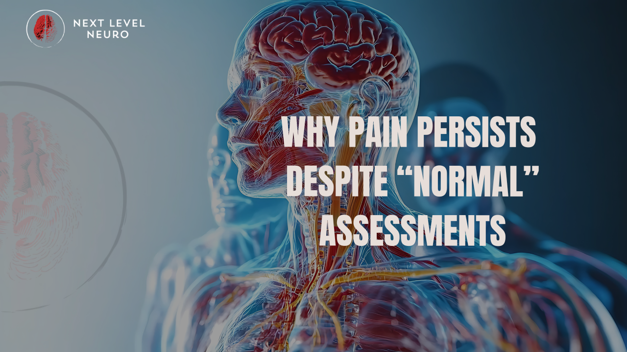
If your imaging is normal, why does the pain still exist?
The Question This Article Answers
Why does pain continue when imaging shows no structural damage and tissues appear healed?
Direct Answer
Pain can persist despite normal imaging because pain is not a direct readout of tissue damage. It is an output of the nervous system. When the brain perceives uncertainty, threat, or unreliable sensory information, it may maintain pain as a protective response—even in the absence of current injury. This decision is influenced by sensory input quality, prior injury history, stress load, movement confidence, and perceived safety, not solely by structural findings on scans.
If your imaging is normal, why does the pain still exist?
That question sits underneath more frustration than most people realize.
Because when an MRI is clean, the expectation is simple:
Pain should stop.
Movement should feel safe again.
Confidence should return.
And when it doesn’t, people start questioning themselves.
Clinicians start questioning the plan.
And the conversation quietly stalls.
This article answers a specific question:
Why can pain persist even when imaging shows no structural damage?
Not in theory.
Not philosophically.
But in the way the nervous system actually works.
To answer that, we have to separate what imaging can show
from what pain actually represents.
One of the most confusing moments for people in pain sounds like this:
“Your MRI looks fine.”
No disc herniation.
No tear.
No structural damage that explains the symptoms.
And yet, the pain is still there.
Sometimes worse.
Sometimes louder.
Sometimes spreading.
This is the moment many people start doubting themselves.
And many clinicians start running out of answers.
But this situation isn’t mysterious when you understand how pain actually works.
Imaging Shows Structure — Not Interpretation
X-rays, MRIs, and scans are excellent at showing tissue structure.
They can tell us:
- If a bone is fractured
- If a disc is bulging
- If there is visible degeneration or tearing
What they cannot show is how the brain is interpreting the body.
Pain is not produced by tissues alone.
Pain is produced by the nervous system’s decision-making process.
And that decision is influenced by far more than structure.
Pain Is an Output, Not a Damage Report
Pain is the brain’s way of saying:
“Something here doesn’t feel safe.”
That “something” might be tissue damage.
But it can also be:
- Threat
- Uncertainty
- Poor sensory input
- Past injury history
- Stress load
- Loss of movement confidence
- Dysregulated autonomic tone
In other words, pain is not a scan result.
It’s a protective output.
When imaging is normal and pain persists, it usually means the nervous system is operating in a protective state, even though no current damage exists.
Why the Brain Stays Protective
The brain relies on sensory input to decide whether a region of the body is safe to move, load, or trust.
That input comes from:
- Vision
- Balance (vestibular system)
- Proprioception (joint position and movement sense)
- Interoception (internal state awareness)
If that information is:
- Inconsistent
- Blunted
- Threat-associated
- Or historically linked to pain
The brain may keep the “pain alarm” on as a precaution.
This is why people can experience:
- Pain without injury
- Pain long after tissue healing
- Pain that moves or changes quality
- Pain that flares under stress or fatigue
The nervous system has learned that this area is uncertain, even if the structure is intact.
Why “Nothing Is Wrong” Feels So Wrong
Being told “everything looks normal” often IS INTERPRETED AS:
“This must be in your head.”
That’s not accurate and it’s not helpful.
Pain is real.
What we feel is real.
But the driver may no longer be tissue damage.
When the nervous system remains in a heightened protective mode, it can amplify signals, lower thresholds, and interpret normal input as dangerous.
The issue isn’t imaginary.
It’s neurophysiological.
Where Applied Neurology Changes the Conversation
This is where applied neurology offers a different lens.
Instead of asking only:
“What structure is damaged?”
Applied neurology asks:
“What information is the brain using to make this decision?”
It evaluates how the nervous system is:
- Processing sensory input
- Regulating threat and safety
- Coordinating movement
- Adapting under load and stress
And most importantly, it tests and retests those systems in real time to see what actually changes the brain’s output.
Pain reduction often follows clarity.
The Missing Piece for Many People in Pain
If you’ve been told:
- Your scans are clean
- Rehab hasn’t helped
- Strength didn’t fix it
- Rest didn’t solve it
It doesn’t mean you’ve failed treatment.
It usually means the lens was incomplete.
Structure matters, but it’s only part of the equation.
The nervous system decides whether pain persists.
We break this down in detail here:
What Is Applied Neurology?
👉 Read the full explanation on our website
That’s where the complete framework lives.
This NLN Substack post is where we explore Why Does Pain Persist Even When Imaging Is Normal?
If pain has taught you anything, it’s that answers aren’t always where we expect them to be.
Sometimes the scan is clear....
....and the signal is still loud.
That’s not a contradiction.
It’s a clue.
New To Applied Neurology - Where to Start
If this model resonates, the next step is learning how to assess and influence regulation without guessing.
That’s why we built The Neuro Advantage.
It teaches:
-
the Threat Bucket model
-
the Input → Output framework
-
practical ways to test and reassess regulation
It’s the fastest way to start working with stress and pain as nervous system outputs.
New To Applied Neurology - Where to Start
For a deeper explanation of this approach, see:
Applied Neurology Fundamentals
The Next Level Neuro Mentorship
FAQ: Pain With Normal Imaging
Can pain be real if imaging is normal?
Yes. Pain is generated by the nervous system, not by imaging findings. Scans show tissue structure, but they do not show how the brain is interpreting sensory input, threat, or safety. Pain can persist even when tissues appear normal if the nervous system remains in a protective state.
Does normal imaging mean the pain is “in my head”?
No. Pain is a real physiological experience. When imaging is normal, it means the driver is likely neurological rather than structural. This includes sensory processing, threat perception, stress load, and movement confidence, not imagination or exaggeration.
Why does pain last long after an injury has healed?
Tissues heal faster than the nervous system relearns safety. After an injury, the brain may maintain protective patterns such as increased sensitivity, muscle guarding, or altered movement. If those patterns are not addressed, pain can persist even after tissue healing is complete.
Why does stress make pain worse when there’s no injury?
Stress increases nervous system vigilance. Under stress, the brain lowers pain thresholds, increases muscle tone, and becomes more protective. This can amplify pain signals even without tissue damage because the nervous system prioritizes safety during perceived threat.
Why doesn’t strength training always resolve pain?
Strength improves capacity, but pain is influenced by safety perception. If the nervous system does not feel safe, strength gains may not integrate properly. Without addressing sensory input, coordination, and regulation, pain can persist despite increased strength.
What role do sensory systems play in persistent pain?
Sensory systems such as vision, vestibular function, proprioception, and interoception inform the brain about safety. When these systems provide unclear or inconsistent input, the brain may increase protection through pain, stiffness, or movement restriction.
How does applied neurology approach pain differently?
Applied neurology evaluates how the nervous system processes information and produces outputs like pain or movement limitation. It uses real-time testing to determine which sensory or neurological inputs change pain immediately, guiding targeted interventions rather than relying solely on structure.
Can applied neurology help even if imaging shows degeneration?
Yes. Structural findings like degeneration are common and often present without pain. Applied neurology helps determine whether those findings are driving symptoms or whether the nervous system is the primary factor maintaining pain.
Is chronic pain permanent if imaging is normal?
Not necessarily. Chronic pain reflects learned protective patterns within the nervous system. With the right inputs, context, and regulation strategies, the nervous system can adapt and reduce pain over time.
What is the difference between pain and injury?
Injury refers to tissue damage. Pain is the nervous system’s protective output. While injury can cause pain, pain can exist without current injury if the brain perceives threat or uncertainty.
Where should I start if my scans are normal but pain continues?
Start by broadening the assessment beyond structure. Understanding how your nervous system interprets movement, stress, and sensory input is often the missing piece when traditional imaging and treatment approaches fall short.
For a full explanation of this framework, see:
👉 What Is Applied Neurology?
Want more information on our Mentorship
and Programs?
We hate SPAM. We will never sell your information, for any reason.
